All living organisms need energy to survive. This energy is provided by small molecular powerplants within the cells of our body – the mitochondria. These organelles have a unique structural design to carry out this essential task: They consist of a smooth outer membrane and a highly folded inner membrane. However, the mechanisms by which the inner membrane adapts its unique shape, has largely remained a mystery until now. A team of scientists led by Stefan Jakobs at the University Medical Center Göttingen and the Max Planck Institute (MPI) for Biophysical Chemistry in Göttingen has now gained new insights into this process by using state-of-the-art microscopy techniques. Since many diseases of the nervous system and the muscular apparatus are associated with a disturbed mitochondrial architecture, the researchers' findings could contribute to a better understanding of how these diseases develop.
The invaginations of the mitochondrial inner membrane, the so-called cristae, house tiny molecular machines that provide energy for our body. “Although the unique inner membrane architecture of mitochondria has been known for decades, it has remained unclear how it forms,” explained Stefan Jakobs, head of the research group Structure and Dynamics of Mitochondria at the MPI for Biophysical Chemistry. The internal structure of a mitochondrion is controlled by a group of proteins known as mitochondrial contact site and cristae organization system (MICOS). Anchored in the inner membrane, MICOS can bend the mitochondrial inner membrane inwards and thus influences its shape. “The individual MICOS components have been known for quite some time,” said Till Stephan, a postdoctoral researcher in the Jakobs group. “But how exactly they interact and how they control the formation of the cristae has been the subject of conflicting ideas.”
To study how the MICOS proteins perform their task, scientists from six research groups at the Göttingen Max Planck Institutes for Biophysical Chemistry and for Experimental Medicine, the University Medical Center Göttingen, as well as the University of Bielefeld worked closely together. In a first step, the researchers used CRISPR/Cas9 to create genetically modified cells in which they could switch the production of individual MICOS components on and off by adding a chemical substance. “Switching off some MICOS components resulted in mitochondria that were no longer able to form cristae correctly,” Jakobs pointed out. The scientists then switched the production of the MICOS proteins on again and investigated how the formation of the MICOS complex from its individual components controlled cristae folding.
Combining fluorescence microscopy and 3D electron microscopy
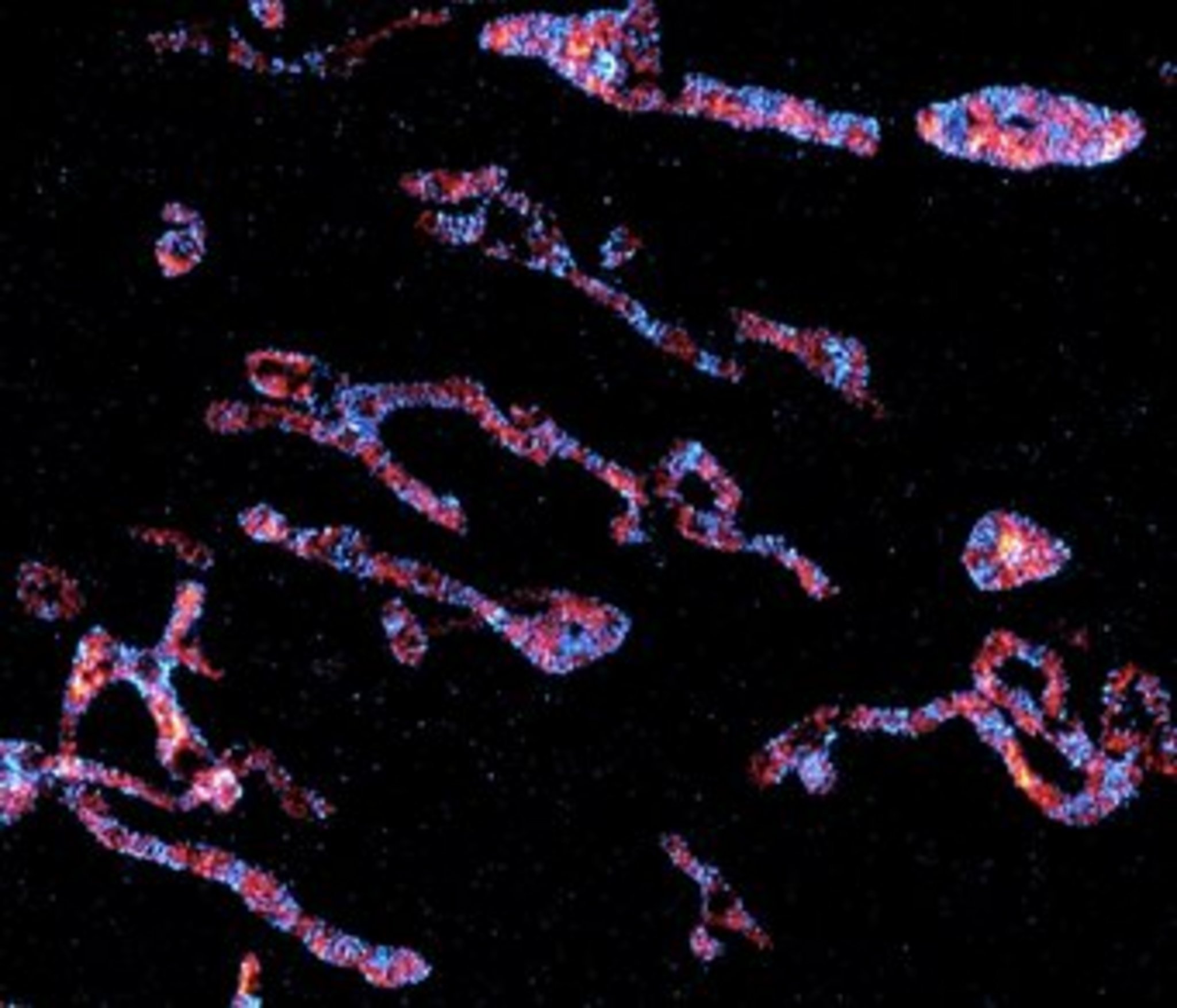
To do so, they relied on 3D electron microscopy and ultra-high resolution fluorescence light microscopy. “A single microscopy method would not have helped us to observe these processes,” the cell biologist emphasized. “With fluorescence imaging methods we can visualize the proteins with nanometer precision. However, to make the membrane visible in which these proteins are located, we needed another approach.” Christian Brüser, a postdoctoral fellow, added: “Only the combination of data from fluorescence microscopy and reconstructions of the mitochondrial membranes based on 3D electron microscopy revealed how the individual proteins work together to form the cristae.”
MICOS consists of two parts, one of which anchors the invaginations of the inner membrane. If this first part then recruits the second part of MICOS, a molecular switch is flipped that controls the inner membrane remodeling. During this process, larger membranes are broken down into smaller pieces to form the numerous, highly ordered cristae within a mitochondrion.
In the future, the researchers hope to work closely with physicians from the University Medical Center Göttingen to further investigate the role of aberrant cristae formation in the development of Parkinson's and Alzheimer's diseases.
Contact
Prof. Dr. Stefan Jakobs
Research Group Leader Structure and Dynamics of Mitochondria at the MPI for Biophysical Chemistry and Professor at the University of Göttingen
+49 551 201-2531
sjakobs@...
Dr. Carmen Rotte
Press Officer and Head of Public Relations
+49 55 1201-1304
carmen.rotte@...
Original Publication
Till Stephan, Christian Brüser, Markus Deckers, Anna M Steyer, Francisco Balzarotti, Mariam Barbot, Tiana S Behr, Gudrun Heim, Wolfgang Hübner, Peter Ilgen, Felix Lange, David Pacheu‐Grau, Jasmin K Pape, Stefan Stoldt, Thomas Huser, Stefan W Hell, Wiebke Möbius, Peter Rehling, Dietmar Riedel, Stefan Jakobs
MICOS assembly controls mitochondrial inner membrane remodeling and crista junction redistribution to mediate cristae formation. The Embo Journal.
Source | DOI

